Optical coherence tomography (OCT)
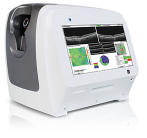
We just added the new Optovue iScan OCT to help with assessing the health of your eyes. Similar to an MRI or CT scan for the eye, the i-Scan OCT uses the SD-OCT to produce cross sectional images of the retinal layers – which is sensitive area of the back of your eye – in a matter of seconds.
Many eye problems can develop without warning and progress without symptoms. In early stages, you may not even notice a change in your vision. But sight threatening conditions and diseases such as macular degeneration, glaucoma, diabetic retinopathy and others can be detected with a thorough exam of the retina. |
Color Fundus Photography
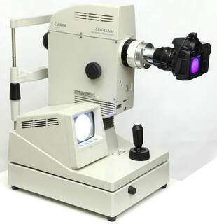
Retinal photography uses a fundus camera to record color images of the condition of the interior surface of the eye also including the retina, retinal vasculature, optic disc, macula, and posterior pole (i.e. the fundus) in order to document the presence of disorders and monitor their change over time.
|


